#IT & Technology - Telecom
Software Enhances Super-res Microscopy
Researchers using microscopy for cell and tissue imaging can now obtain optical resolution of up to 120 nm thanks to a software upgrade from Olympus
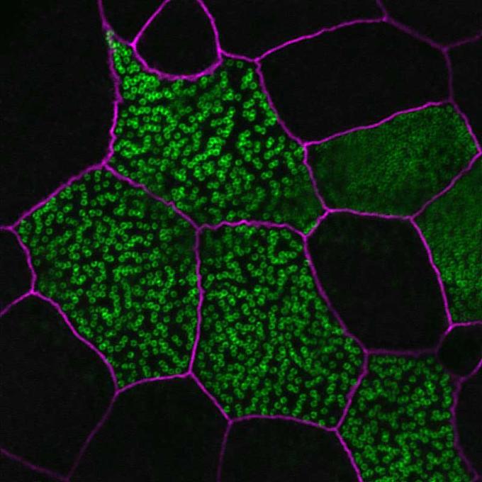
With Olympus FV-OSR software, the FluoView FV1200 confocal laser scanning microscope can be transformed into a powerful system for super resolution microscopy. Introducing a smart route to super resolution, the software module enables FV1200 microscope users easy access to what was previously the sole domain of highly specialized microscopy systems. The extra detail offered by super resolution technology helps researchers reveal the subtlest mechanisms at work within cells or tissue. Although three-dimensional confocal laser scanning microscopy achieves vastly improved lateral and axial resolution compared to standard widefield imaging, it is still limited by the diffraction limit of light.
In certain settings, images can greatly benefit from enhanced contrast after post-acquisition processing. This is where super resolution microscopy comes into play, breaking the diffraction limit of light and enhancing cell- and tissue-imaging studies. The software controls specific hardware settings and advanced signal processing. Operation has been designed with users in mind, and requires only minimal training to achieve expert results. Capitalizing on the flexibility of the modular IX83 automated inverted microscope platform, the FV1200 system is optimized for a diverse array of live cell imaging applications, including fluorescence recovery after photobleaching (FRAP), fluorescence loss in photobleaching (FLIP) and photoactivation.
